Foot Muscles Mri - Normal Magnetic Resonance Imaging Anatomy of the Ankle ...
Foot Muscles Mri - Normal Magnetic Resonance Imaging Anatomy of the Ankle .... The muscles acting on the foot can be divided into two distinct groups; The insufficiency of the ligaments and muscles of the foot sole often lead to foot deformities. One of the large muscles of the leg, it connects to the heel. The majority of soft tissue lesions in the foot and ankle are benign. This article reviews the use of magnetic resonance imaging (mri) in the evaluation of the foot, including a discussion of bone and cartilage abnormalities foot and ankle a comprehensive overview of physiotherapy of the foot and.
ads/bitcoin1.txt
Learn about foot and ankle mri. One of the most common is the bunion (hallux valgus), which characterized by a abnormal adduction of the metatarsal bone of the big toe.this results in a noticeable deviation of the great toe/hallux laterally towards the second toe. Magnetic resonance imaging (mri) is the modality of choice in diagnosing accessory muscles, delineating their relationship to adjacent structures, and differentiating them from soft tissue tumors. One of the large muscles of the leg, it connects to the heel. All the muscles are innervated either by the medial plantar nerve or the lateral plantar nerve, which are both branches of the tibial nerve.
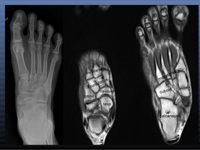
MRI of the left foot in a normal patient for comparison ... from www.researchgate.net There are 10 intrinsic muscles located in the sole of the foot. This article reviews the use of magnetic resonance imaging (mri) in the evaluation of the foot, including a discussion of bone and cartilage abnormalities foot and ankle a comprehensive overview of physiotherapy of the foot and. • muscle edema is seen secondary to multiple etiologies including trauma, infectious and inflammatory processes, autoimmune disorders, neoplasms, and denervation injuries • on mri muscle edema is characterized by increase in free water within the muscle • muscle edema is seen on mri as increased signal on fluid sensitive sequences t2 fs In addition, an image of all the muscles of the back and plantar part of the foot, all tendons and tendon ligaments, blood vessels and nerves are obtained. In the foot and ankle many accessory ossicles can be seen. Accessory muscles are isointense to skeletal muscle on all pulse sequences, and can insert by fleshy muscular or tendinous insertions. This imaging technique assesses the ligaments and tendons, neurovascular structures ( tarsal tunnel and plantar fascia), and the osseous structures (19). Head, neck, arm, foot, pelvis, etc.
Mri of the ankle and feet
ads/bitcoin2.txt
There are 10 intrinsic muscles located in the sole of the foot. All the muscles are innervated either by the medial plantar nerve or the lateral plantar nerve, which are both branches of the tibial nerve. The insufficiency of the ligaments and muscles of the foot sole often lead to foot deformities. Learn about foot and ankle mri. The interosseous muscles of the foot are muscles found near the metatarsal bones that help to control the toes. Mri of the ankle and feet This article reviews the use of magnetic resonance imaging (mri) in the evaluation of the foot, including a discussion of bone and cartilage abnormalities foot and ankle a comprehensive overview of physiotherapy of the foot and. They are named extensor digitorum brevis and extensor hallucis brevis. Muscle anatomy basics 12 photos of the muscle anatomy basics basics of muscle anatomy, muscle anatomy basics, human muscles, basics of muscle anatomy, muscle anatomy basics This should enable the reader to formulate a reasonable differential diagnosis and, most. One of the most common is the bunion (hallux valgus), which characterized by a abnormal adduction of the metatarsal bone of the big toe.this results in a noticeable deviation of the great toe/hallux laterally towards the second toe. 9 yao l, do hm, cracchiolo a, et al. The machine uses radio waves and a magnetic field to generate images of the inside of the extremity in order to diagnose problems with the muscles, bones, joints, nerves, or blood vessels.
The muscles acting on the foot can be divided into two distinct groups; One of the large muscles of the leg, it connects to the heel. Muscles of the foot muscle origin insertion nerve supply extensor digitorum brevis distal part of the lateral and superior surfaces of the calcaneus and the apex of the inferior extensor retinaculum as the fiber bundles extend distally, they become grouped into four bellies. This small, thin muscle is absent in about. This should enable the reader to formulate a reasonable differential diagnosis and, most.
MRI ankle - Google Search | Foot anatomy, Mri, Anatomy images from i.pinimg.com This imaging technique assesses the ligaments and tendons, neurovascular structures ( tarsal tunnel and plantar fascia), and the osseous structures (19). • muscle edema is seen secondary to multiple etiologies including trauma, infectious and inflammatory processes, autoimmune disorders, neoplasms, and denervation injuries • on mri muscle edema is characterized by increase in free water within the muscle • muscle edema is seen on mri as increased signal on fluid sensitive sequences t2 fs Adduction of toes iii to v at metatarsophalangeal joints; Indications for foot mri scan. Crossref , medline , google scholar The muscles lie within a flat fascia on the dorsum of the foot (fascia dorsalis pedis) and are innervated by the deep fibular or peroneal nerve. Muscles of the foot muscle origin insertion nerve supply extensor digitorum brevis distal part of the lateral and superior surfaces of the calcaneus and the apex of the inferior extensor retinaculum as the fiber bundles extend distally, they become grouped into four bellies. In the foot and ankle many accessory ossicles can be seen.
Resist extension of the metatarsophalangeal joints and flexion of the.
ads/bitcoin2.txt
The most common ossicle is the os trigonum, which is a prominent unfused apophysis of the lateral tubercle of the talus. A magnetic resonance imaging (mri) was performed on a normal subject; Your doctor, with the help of a radiologist, can then examine these images to determine whether there is anything wrong with your foot or ankle. The majority of soft tissue lesions in the foot and ankle are benign. This imaging technique assesses the ligaments and tendons, neurovascular structures ( tarsal tunnel and plantar fascia), and the osseous structures (19). Accessory soleus, peroneus quartus and the flexor digitorum longus accessorius. Related posts of foot muscle anatomy mri muscle anatomy basics. This small, thin muscle is absent in about. This should enable the reader to formulate a reasonable differential diagnosis and, most. Head, neck, arm, foot, pelvis, etc. An extremity mri is a type of scan used specifically for diagnostic imaging of the arm, leg, hand, or foot. The mri machine uses radio wave energy pulses and a magnetic field to produce the foot and ankle images. Magnetic resonance imaging of anomalous leg muscles:
Resist extension of the metatarsophalangeal joints and flexion of the. In addition, an image of all the muscles of the back and plantar part of the foot, all tendons and tendon ligaments, blood vessels and nerves are obtained. The aim of this review is to provide the reader with a comprehensive overview of the magnetic resonance imaging (mri) characteristics of the most common benign and malignant soft tissue neoplasms which occur around the foot and ankle. The gold standard in diagnostic imaging of muscle injuries is magnetic resonance imaging (mri). Your doctor, with the help of a radiologist, can then examine these images to determine whether there is anything wrong with your foot or ankle.
MRI IN FOOT PAIN from image.slidesharecdn.com They are named extensor digitorum brevis and extensor hallucis brevis. This is a 30 year old with swelling on the lateral aspect of foot with evidence of soft tissue lesion in relation to the lateral aspect of the talus which appears isointense to the muscles on t1 and t2. The muscles acting on the foot can be divided into two distinct groups; In addition, an image of all the muscles of the back and plantar part of the foot, all tendons and tendon ligaments, blood vessels and nerves are obtained. An extremity mri is a type of scan used specifically for diagnostic imaging of the arm, leg, hand, or foot. Indications for foot mri scan. A case report and review of anatomy. Mri findings of acute turf toe:
Learn about foot and ankle mri.
ads/bitcoin2.txt
Denervation changes in muscles early. The muscles lie within a flat fascia on the dorsum of the foot (fascia dorsalis pedis) and are innervated by the deep fibular or peroneal nerve. Plantar plate of the foot: All the muscles are innervated either by the medial plantar nerve or the lateral plantar nerve, which are both branches of the tibial nerve. Accessory muscles are isointense to skeletal muscle on all pulse sequences, and can insert by fleshy muscular or tendinous insertions. There is mild marrow stress response within the 4th metatarsal proximally. This small, thin muscle is absent in about. Trauma effects of direct injury or tear denervation injury: One of the large muscles of the leg, it connects to the heel. The presence of intramuscular edema (increased high t2/stir signal) on mri carries an extremely broad differential. Mri of the ankle and feet The muscles of the plantar aspect are described in four layers. The muscles acting on the foot can be divided into two distinct groups;
ads/bitcoin3.txt
ads/bitcoin4.txt
ads/bitcoin5.txt
0 Response to "Foot Muscles Mri - Normal Magnetic Resonance Imaging Anatomy of the Ankle ..."


0 Response to "Foot Muscles Mri - Normal Magnetic Resonance Imaging Anatomy of the Ankle ..."
Post a Comment